
Select Page
Study Shows Intermittent Fasting Did Not Reduce Weight More Than Regular Eating

Study Shows Intermittent Fasting Did Not Reduce Weight More Than Regular Eating

Study Shows Intermittent Fasting Did Not Reduce Weight More Than Regular Eating

October 23rd, 2020

  • A 12-week study compared the weight loss of two groups; 1 group fasted for 16 hours of the day, while the other group ate throughout the day without time restrictions
  • The clinical trial included 141 adults, the largest randomized, controlled study for intermittent fasting
  • Intermittent fasting did not statistically improve weight loss (or other body composition measurements) compared to normal eating

Intermittent fasting, also referred to as time restricted eating (TRE), has recently gained traction as an easy and effective way to lose weight. It consists of eating within a restricted time frame and fasting for several hours in between. However, most of the benefits associated with it are anecdotal, and are not based on research that involves a control arm.

Side note: The most conclusive clinical trials include design protocols with a control arm, randomization, and a large enough sample set. Unlike the majority of other fasting trials that lack these designs, this study included all of these protocols.

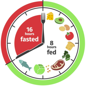
A randomized clinical trial conducted by a group in California was testing the effects of TRE on weight and other metabolic parameters in overweight and obese individuals. They hypothesized that those restricted to an 8-hour eating period (image below) would lose weight and improve other metabolic parameters as compared to those eating 3 meals a day without timing restrictions, also referred to as consistent meal timing (CMT).
A total of 141 participants were randomized between the two interventions (TRE or CMT) and 105 completed the entire protocol. The study lasted 12 weeks (~3 months). The TRE group was not told what or how much to eat, they were only restricted to the time of eating. Between 12:00 PM to 8:00 PM, they were allowed to eat to their desire. Then from 8:00 PM until 12:00 PM the following day, they were instructed to fast. The only thing permitted during their fasting hours were non-caloric beverages.

What the Data Showed:

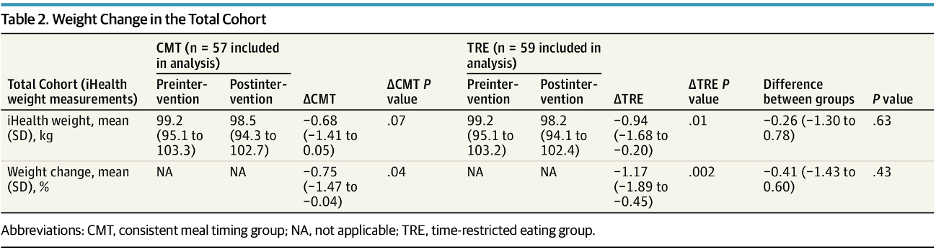
In the consistent meal timing (CMT) arm, participants lost an average of 0.68Kg over the 12-week period, equivalent to 0.75% decline in body weight. In the time restricted eating (TRE) arm, participants lost an average 0.94Kg, or approximately 1.17% decline in body weight. See table below.

Comparing the difference between the two groups (0.94kg – 0.68kg = 0.26kg), the p-value was 0.63. This denotes that the difference is not a statistically significant change, and thus can be a result of chance. In simple terms, time restricted eating did not cause the drop in weight and was no different than consistent meal eating.
It was also found that there was no significant change in the body fat mass in the TRE or CMT group. There was a decrease in lean mass and total energy expenditure but no change in respiratory quotient, resting metabolic rate, fasting glucose, fasting insulin, triglycerides, LDL, HDL or total cholesterol.

In short, in this study, TRE did not statistically improve any body composition measurements.

Arguments can be made that a 12-week time period may not be adequate to see the lasting metabolic effects. A longer-term study, say 24-weeks, could be more telling on how the body will be rewired during a metabolic change. Additionally, a further limitation of this study can be that the amount of food was not restricted, only the timing was. Calories in & out is still an important feature of any diet plan. 

TRE Led To Lean Mass Loss, More Than Fat Loss

It is important to mention the results found regarding lean mass. A proportion of the participants (n=50) in the California study were tested in person, while the remainder of them recorded their data using a custom app.

Of the ones that went in person, there was an average weight loss of 1.70 kg. Of this total weight, 1.10 kg was lean mass while the remaining weight was fat mass. Usually, 20-30% of weight loss is lean mass so the 1.10 kg of lean mass lost was far more than that (~65%).

It has been seen that calorie and protein intake is reduced with a TRE diet. This can be a problem for those with sarcopenia (degenerative muscle loss) or even those who are trying to maintain their muscle mass while losing weight as there is a well-defined relationship between protein intake and muscle mass.

Thus, it is important to maintain a high protein diet while intermittent fasting to avoid the loss in lean mass.

Other Studies Reached Different Conclusions

Results found in this study were inconsistent with other reports claiming that TRE is beneficial for weight loss and other metabolic parameters.

One study by Jamshed et al., found that TRE improves glucose levels as well as secondary markers like sleep and aging.

Another study showed that TRE lead to a 3% decrease in weight and also improved cardiovascular health. However, it is important to note that the sample size in this study was small (n=19) and no control group was included.

A third study showed that 4 and 6 hour TRE reduced daily intake by 550 calories and led to statistically significant weight loss (3% decline in body weight) over 8 weeks. This was a randomized, controlled study that collected data from 58 participants.

Conclusions to Make

Overall, intermittent fasting is an appealing, fuss-free weight loss alternative. It provides individuals with the freedom to eat as desired, as long as feeding is conserved to restricted hours. Based on the results in this study, there were limited changes between the TRE and CMT groups. Weight loss was not any greater in the TRE group in comparison, and insignificant changes were seen in other metabolic parameters such as fasting glucose and insulin or blood lipids and cholesterol.

This study showed that TRE did not result in substantial weight loss when compared to the CMT group. Also, although protein intake amongst participants was not directly measured in this study, the loss of lean mass seen suggests that protein levels are lowered in a TRE diet.

In the absence of other interventions such as reduced caloric intake, timing of fasting hours (early vs late) or increased levels of protein, TRE has not shown to be a more effective option of losing weight. It would be of interest to future studies to further explore the relationship between these interventions alongside TRE for weight loss.
NK Cells: Oncology’s Next Big Wave

NK Cells: Oncology’s Next Big Wave

NK Cells: Oncology’s Next Big Wave

October 18th, 2020

Key Points:

  • Although two CAR-T therapies have received FDA approval, shortcomings limit their usability in a real-life setting
  • Allogeneic “off-the-shelf” CAR-T approaches also face two main hurdles: GvHD and graft rejection
  • NK therapies, including engagers and CAR-NK, have seen a recent spike in interest due to their superior safety potential and broad manufacturing capabilities
  • Next-gen cell therapy approaches are discussed, include allogeneic CAR-T & CAR-NK, NK cell engagers and the different NK cell sources

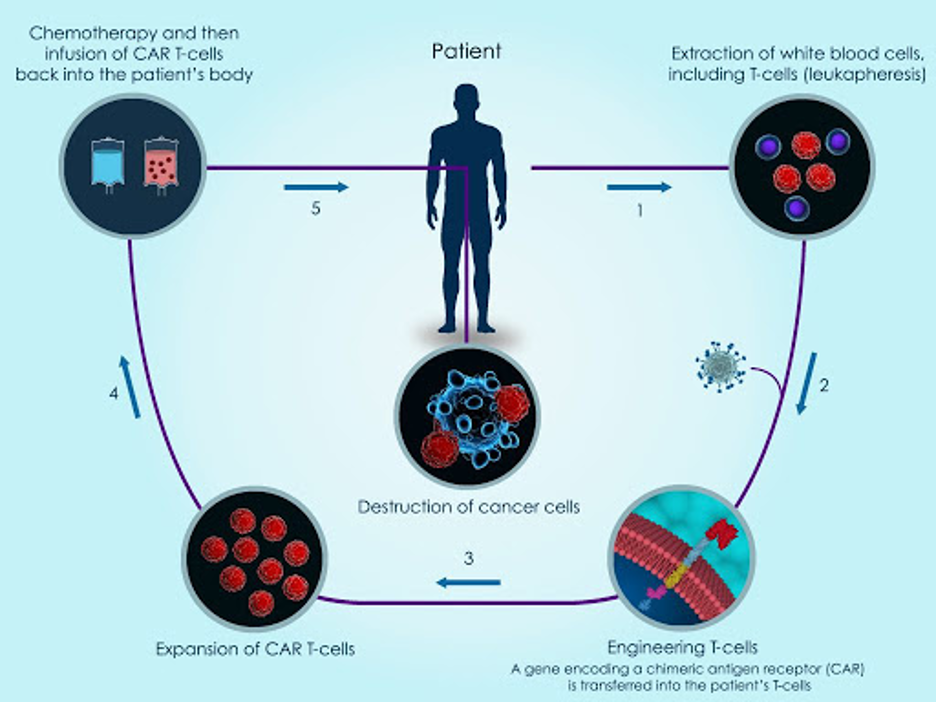
For several years, chimeric antigen receptor T-cell (CAR-T) therapy has been the topic of discussion as the next-gen treatment for oncology. CAR-T works by using T cells from the patient’s own blood, referred to as “autologous cells”. The T cells are collected, genetically modified and multiplied, and then re-injected back into the patient (see image below).

The idea behind CAR-T cells is that a synthetic receptor is attached to the T cells and when this receptor comes into contact with the antigen of an infected cell, intracellular domains activate the cytotoxicity of the T cell. In simple terms, T cells are genetically modified and enhanced to better detect cancer cells when they are reinjected back into the body.

CAR-T Shortcomings Limit Usability in Real Life Setting

Two CAR-T cell therapies have been approved by the FDA, both showing promising results. However, they come with their own set of challenges. Most people suffering with cancer, who could benefit from the therapy, are not receiving it. This is because it involves a lengthy manufacturing process with a high cost profile and could have potentially severe side effects.

Allogeneic Approach Has Answers to Existing CAR-T Limitations, But Other Issues Arise

To make CAR-T more accessible to those in need, researchers are using T cells extracted from a third-party donor (called “allogeneic”). This off-the-shelf concept is similar to the autologous method discussed above but allows for the cells to be easily accessible in the hospital setting. This way, large proportions of pre-engineered cells can be preserved for use when needed and one donor’s T cells can be administered to several patients, thus lowering the manufacturing costs and time.

There are two main hurdles with the off-the-shelf approach:

  1. Graft-versus-host disease (GvHD) – Occurs when the donor T cells view the patient’s cells as foreign and attack them, causing serious or fatal side effects.
  2. Graft rejection – Graft rejection is the opposite process. The patient’s own immune system detects the donor T cells as foreign and rapidly eliminates them, thereby limiting their anti-tumor activity.

Modifications Helping with Allogeneic CAR-T Hurdles

Cellectis and Allogene are two companies making off-the-shelf CAR-T cells genetically engineered to avoid GvHD and graft rejection. Using Cellectis’ gene editing technology, Allogene makes two modifications:

  1. Eliminate functional T cell receptors (TCRs) that prevent the CAR-T cell from recognizing a patient’s normal tissue cells as foreign. Without these TCRs, the CAR-T cells cannot bind to healthy cells found in the patient. This is meant to improve safety and limit the risk of GvHD.
  2. Knocks out the CD52 surface protein on the CAR-T cells. In addition, Allogene uses their own anti-CD52 monoclonal antibody to suppress the host immune system from recognizing CD52 on the surface of foreign CAR-T cells. Without CD52 on CAR-T & use of anti-CD52 antibody, Allogene believes that the CAR-T & patient immune system will not be attacking each other. Instead, both will work together to attack the tumor cells.

See a visual representation of the two modifications Allogene has made to control GvHD & graft rejection (below). So far in early Phase 1 studies, Allogene has not reported any cases of GvHD or graft rejection in 22 patients.

The NK Cell Approach to Treating Cancer

The above-mentioned CAR-T limitations have sparked a recent interest in NK cells as an alternate treatment approach.  Early studies were focused on infusing non-targeted endogenous NK cells back into patients (non-targeted NK cells do not have any antibodies or modifications). This resulted in a lack of tumor recognition by autologous NK cells. Therefore, it was determined that a combination of NK cells + target therapies was needed.

Side Note: What differentiates NK cells from their lymphocytic counterpart T cells is their ability to kill cancer cells without sensitization. Yet, when primed with cytokines, the cytotoxic function of NK cells is increased along with their longevity in vivo.

To get a better understanding of NK cells and their basic biology, read LifeScite’s NK Cells for Dummies: Understanding the Next Cancer Paradigm article.

Combinations With NK-Cell Engagers

To improve NK cell antitumor activity, pharma companies are combining antibody targeting agents with NK cells. Affimed recently entered Phase 1 trials that will test a combination of NK cell engager AFM13 with MD Anderson’s off-the-shelf NK cells.

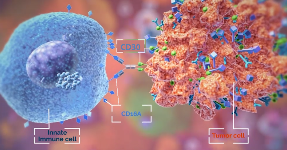
AFM13 is an antibody which binds to CD16A receptor found on NK cells and other innate immune cells like mast cells, macrophages and monocytes. The other end of the antibody will target the CD30 antigen expressed on lymphoma tumor cells (see image below). The hope is that NK cells equipped with better targeting agents will lead to added antitumor activity and therefore superior efficacy.

Similarly, Innate Pharma’s monoclonal antibody monalizumab blocks the checkpoint NKG2A receptor found on NK & certain T cells. By blocking this inhibitory receptor, Innate believes that the cancer fighting cells (NK cells and T cells) will be able to better detect, and bind, the HLA-E receptor found on tumorous cells. The company is advancing their innate checkpoint blocker into a Phase 3 study in combination with cetuximab.

CAR-NK Do Not Share CAR-T Limitations

Similar to CAR-T, CAR-NK cells are genetically equipped with receptors on their surface which seek signs of danger or stress that might indicate a tumor, or a cell infected with a virus. When they find such a cell, NK cells can attack it directly and call T cells to the site to help.

Data thus far shows that CAR-NK cells contain several benefits over their CAR-T cell counterparts:

  1. Superior Safety Profile: CAR-NK cells have a low risk of cytokine release syndrome compared to CAR-T cells because they secrete restricted levels of pro-inflammatory cytokines IFN-γ, IL-1 and IL-6.
  2. Broad Production Potential: Low likelihood of triggering GvHD upon allogeneic infusion means that CAR-NK cells may be prepared as an “off-the-shelf” product and are not restricted to autologous cells.
  3. CAR-NK cells can work in CAR-dependent and CAR-independent manners. (Recall that CAR stands for chimeric antigen receptor.)
    • CAR-dependent: Cancer cells are smart and can make themselves “invisible” to immune cells. The CAR added onto NK cells now gives them the ability to recognize and kill a target on cancer cells that were once “invisible”.
    • CAR-independent: CAR-NK cells also contain native receptors and retain their intrinsic ability to recognize and kill cancer cells independent of CARs. This is because they do not rely on antigen-specific binding to be turned on but rather, can induce a positive or negative signal through binding with ligands on target cells.

NK Cell Sources: Not All NKs Are Equal

NK cells demonstrate promising potential to be prepared as an off-the-shelf product. This, however, requires a donor source where ample, functional NK cells may be harvested. To date, NK cell isolates have most often been extracted from the donor’s peripheral blood (PB-NK cells) or umbilical cord blood (UCB-NK cells).

Obtaining an abundant number of functionally active NK cells from these sources has proven to be difficult. Other and more accessible cell sources include NK-92 cells and stem cell-derived NK cells.

Below is be a breakdown of each of the cell sources.

Peripheral blood (PB-NK cells)

These NK cells are collected from the blood of screened healthy donors. The NK cells (and specific types of T cells) are isolated from the collected blood sample, expanded, and genetically modified with editing technologies like CRISPR to improve persistence and overcome resistance.

Editas Medicine in Cambridge, Massachusetts, is using CRISPR to remove a receptor on NK cells that bind TGF-β — a signaling molecule produced by some tumors that can shut down immune cells. The hope is that removing the receptor will make the CAR-NK cells more potent tumor-killing machines.

Umbilical Cord blood NK Cells (UCB-NK cells) 

NK cells isolated from umbilical cord blood have shown to possess reduced cytotoxicity when compared to peripheral blood derived NK cells. However, the use of umbilical cord blood as a source of NK is also limited in the number of NK cells that can be obtained, when compared to peripheral blood as a source.

A study out of MD Anderson (Rezvani, Liu et al. 2018) collected UCB-NK cells and engineered them with CARs that allowed the innate cells to target the CD19 protein found on select tumor cells. The modified NK cells were also made to express IL-15, a cytokine that activates NK cells, as well as inducible caspase-9 (iC9) as a safety switch. Data (found here) showed early efficacy in several lymphoma cancers and a clean safety profile. This caught the attention of big pharma company Takeda, which licensed the program in late 2019. Takeda estimates that the program can enter pivotal trials in specific refractory lymphoma indications as early as 2021 and have a BLA filing by 2023.

NK Cell Lines 

A specific strand of NK cells called NK-92 are sourced from cancer patients. These withdrawn cells are irradiated so that the cancerous cells are wiped out, and only the functional NK-92 cells remain. After irradiation, NK cell lines are expanded indefinitely, and can be genetically modified to recognize specific antigens found on tumor cells. Various clinical trials with CAR-NK cells use NK92 cell line because of their ability to be expanded indefinitely in vitro and are able to undergo repeated freeze/thaw cycles needed during manufacturing process. This gives NK cell lines ideal features as off-the-shelf products.

However, because NK-92 cells are sourced from a tumor cell line they have several drawbacks including (1) potential tumorigenicity risk (ability to form into tumorous cells) (2) lack of CD16 and NKp44 expression (non-targeted to attack tumors) and (3) loss of in vivo expansion potential due to the irradiation process.

Nantkwest is developing several modified NK-92 cell lines to target cancer. Their t-haNK platform incorporates receptors (CD16, CD19, NKG2D etc.) as well as CAR to target cancer cells and enhance cell killing capabilities.

Stem cell-derived NK cells

Think of human induced pluripotent stem cell (iPSC) as a blank slate. These stem cells can be modified to become 200+ cell types, including NK cells. Stem cell-derived NK cells have the ability to produce unlimited cells for use in the clinic. Their ability to be genetically engineered and unlimited ability to self-renew gives them the potential to generate an infinite number of cells for off-the-shelf CAR-NK cells.

Fate Therapeutics has a pipeline of genetically modified iPSC derived NK cells, that include CD16 to augment antibody therapy, IL-15 to enhance NK cell function, CAR to make NK cells more targeted and CD38 knockout to improve safety. Fate’s iPSC-derived NK cells have shown to be safe and well tolerated in Phase 1 data. Fate signed a collaboration with Johnson & Johnson’s pharmaceutical arm Janssen to build novel off-the-shelf iPSC-derived CAR-NK and CAR-T candidates.

Current State of NK: Early, But Promising

It is clear that NK cells as therapy prove to be a great option for use in the clinic. They provide effective results while overcoming the GvHD associated with their CAR-T counterparts. However, their safety and efficacy has only been shown towards a handful of cancers. The challenge will be seen when solid tumors are targeted. NK cells are still a relatively new concept in the oncology field and thus, should continue to be studied in order to confirm their long-term effects. With the recent big pharma interest, it seems likely that the resources into NK cells will continue pouring in.

NK Cells for Dummies: Understanding the Next Cancer Paradigm

NK Cells for Dummies: Understanding the Next Cancer Paradigm

NK Cells for Dummies: Understanding the Next Cancer Paradigm

October 2nd, 2020

The growing field of NK cell-mediated immunotherapy first started almost two decades ago when it was found to be a safe and effective treatment method for those with advanced-stage leukemia.

Immunotherapy treats disease by stimulating the body’s immune response. The immune system is made of two segments:

  1. innate immunity: non-specific to one antigen, first line of defense that uses natural killer (NK) cells, among others
  2. adaptive immunity: specific to a single antigen, acquired immunity that uses T cells and B cells to invade foreign pathogens

Until very recently, the majority of pharma research was directed towards the adaptive immune system and activating T cells. FDA approved blockbuster therapies (Opdivo, Keytruda, to name a few) followed this line of thinking. In the last few years, NK cell therapies have received a lot of interest. This article will be discussing the fundamentals of NK cells and a follow-on note will focus on NK therapies in development.

NK Cells Share The Same Cancer Killing Features As T Cells

Think of NK cells as a security system for your body that roam around, looking for abnormal cells (ie. tumor or virally infected cells). NK cells are functionally most similar to a type of T cell called CD8+ T cells.

Both NK cells and CD8+ T cells are considered cytotoxic because they are capable of killing infected and cancerous cells. The difference between the two of them is the way in which they carry out this function.

How do NK Cells differentiate normal cells from abnormal cells? 

On their surface, NK cells express inhibitory receptors (that turn off the NK cell) and activating receptors (that turn on the NK cell).

Normal cells in the body display MHC I molecule (inhibitory) as well as activating ligand (stimulatory).  A net balance between inhibition and activation occurs when recognizing a healthy cell. Thus, the NK cell will be kept OFF (no release of cytotoxins = NO killing of normal cell).

Abnormal cells (ie. tumor/virus cells) only display the activating ligands. They will bind to the activating receptors on NK cells and signal it to turn ON, release cytotoxins and KILL the target cell.

How NK Cells Don’t Go into Killing Overdrive

Inhibitory pathways (immune checkpoints) are essential to balance the killing abilities of immune cells. These immune checkpoints maintain self-tolerance and balance the stimulatory pathways to avoid unnecessary damage of normal cells. Issues arise when this negative regulation is affected because immune cells, including NK cells & T cells, lose this “stop” ability. This can lead to an autoimmune disease.

How NK Cells Cooperate with The Adaptive Immune System

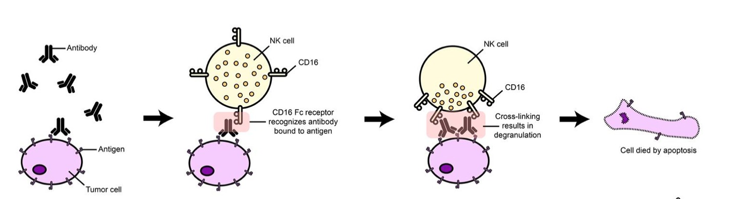
Remember the adaptive immune system includes cancer-killing CD8+ T cells and antibody-producing B cells, among others. Tumor cells express antigens on their surface (see image below). Circulating antibodies of the adaptive immune system (called “IgG”) bind to these antigens and recruit immune cells to the foreign cells.

NK cells are one of the immune cells recruited and this process is called antibody-dependent cell-mediated cytotoxicity (ADCC). The NK cell expresses a receptor called Fc-gamma receptor (FcγR or CD16), which binds to the antibody found on the antigen and leads to its destruction.

NK cells are not defined to one immunity over the other but rather contain functional attributes that pertain to both innate and adaptive immunity. Keep in mind that the methods of NK cell activation mentioned above are an extremely simplified explanation of how NK cells function.

Cancer Cells Can Hide, But for How Much Longer?

Cancer cells are smart and escape detection from the immune system by taking advantage of the immune checkpoints, mentioned above. By increasing their inhibitory ligands that correspond to the inhibitory receptors on immune cells, cancer cells ensure their escape from immune detection.

With T cells, blocking the inhibiting checkpoints with monoclonal antibodies resulted in the blockbuster drugs by Merck (Keytruda) and BMY (Opdivo). PD-L1 is an inhibitory ligand on the surface of cancer cells that binds to PD-1 inhibitory receptors on T cells and inhibits their cytotoxicity. Checkpoint inhibitor drugs use a monoclonal antibody (anti-PD-1) to help T cells restore their ability to detect cancer cells.

A similar approach is being taken with NK cells: targeting inhibitory checkpoints with antibodies in clinical development allows the NK cells to be relieved of inhibition and thus recognize and target cancer cells.

Up to 90% of US COVID Tests Can Be False Positives

Up to 90% of US COVID Tests Can Be False Positives

Up to 90% of US COVID Tests Can Be False Positives

September 21st, 2020

The use of polymerase chain reaction (PCR) testing as a frontline diagnostic tool for COVID is being questioned. These tests provide a positive/negative result for COVID infection, but do not show how much virus a patient has. The amount of virus can make all the difference when it comes to classifying contagious COVID cases.

Testing the RNA sequence of a virus is a complicated task. Even samples infected with the virus do not contain enough of the viral RNA. Thus, amplification is required to multiply and generate enough RNA to be present for adequate testing.

The COVID PCR test uses this exact amplifying mechanism. It involves a series of steps that are repeated over and over, called cycles. These cycles eventually lead to an exponential increase in genetic material (RNA in the case of coronavirus), doubling the amount with each cycle. This means that after 40 cycles (240), there are about 1000 billion copies of RNA.

The problem with PCR testing is that it only provides a yes or no answer, either someone is infected or not. However, it is extremely important to realize that this is a quantitative issue as well. The PCR test does not show how much virus (viral load) someone has.

Simply put, PCR amplifies the RNA sample in a series of cycles. If there is a high viral load, which means the patient is more infectious and contagious, it will be detected in fewer cycles.

PCR Test Sensitivity Greater Than Live COVID Viral Load 

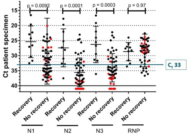
For COVID testing, the appropriate cut-off needed to detect live virus is between 30-34 cycles, as found by a French study. Detecting the virus at <34 cycles indicates that there is enough viral load to be contagious. Detecting the virus at >35 cycles could indicate that there are just genetic fragments of the virus that pose no particular risk (ie. patient has COVID, but is not contagious).

The latter situation has the potential to lead to a high level of false positive test results. Some researchers estimate that this false postive scenario make up to 90% of COVID cases in the US.

Most PCR tests used in the US set the COVID cut-off limit between 37-40 cycles. Remember, the French study above claimed that if a virus sample needs to be amplified more than 34 cycles in order to be detected, then it is not viable and thus the person is neither infected nor contagious. Even the CDC has suggested that it is difficult to detect any live virus in a sample above a threshold of 33 cycles.

There seems to be a discrepancy between the contagious viral amount needed (~34 cycles) and what PCR tests are diagnosing (~40 cycles). This inconsistency has the potential to result in false positives and inflated COVID cases.

The New York Times reported that in three sets of testing data that modified thresholds under 40 cycles, between 43-90% of positive COVID tests carried viral loads that could be considered non-contagious.

Relying Solely on COVID Case Numbers Is Flawed

The current rhetoric relies on media headlining the number of cases to dictate the severite of the COVID situation. Clearly, with over-sensitive testing, the case numbers alone do not tell the full story. In fact, the case numbers are likely to be inflated and overstating the severity of COVID. Alternative data poins, such as hospitalizations and deaths, must be taken into consideration to reach a more accurate conclusion on the state of disease. 

Greater transparency is needed from the manufacturers of PCR tests and labs that oversee the testing process. The FDA, CDC and overall population need to push for more testing, with multiple thresholds and additional data points. A simple “Yes” or “No” is not sufficient, nor should it be the standard.

Trillium’s CD47 Monotherapy Data Draws Attention of Pfizer and Wall Street

Trillium’s CD47 Monotherapy Data Draws Attention of Pfizer and Wall Street

Trillium’s CD47 Monotherapy Data Draws Attention of Pfizer and Wall Street

September 17th, 2020

Last week, Trillium reported topline data from their latest cohort for TTI-622, the CD47 targeting candidate. The monotherapy data triggered a series of events that followed, which included a $25M investment from Pfizer and a $150M financing from healthcare focused Wall Street funds.

Below is why Trillium’s approach is garnering attention from big pharma and Wall Street.

CD47 Approach Is Interesting Due to Broad Tumor Applicability

A large number of hematologic and solid tumors express high levels of CD47, providing a “do not eat me” signal by binding to signal regulatory protein alpha (SIRPα). This “do not eat me signal” prevents innate immune cells (first line of immune defense; NK-cells, macrophages, to name a few) from attacking tumor cells. Think of CD47 as an evasive tool that tumor cells use to stay off the radar of the immune system.

It is believed that a CD47-blocking antibody will enhance the immune system’s ability to detect tumors that are currently evading the immune system. The rapidly evolving CD47 space has seen an increase in interest recently with big money and big pharma allocating research dollars.

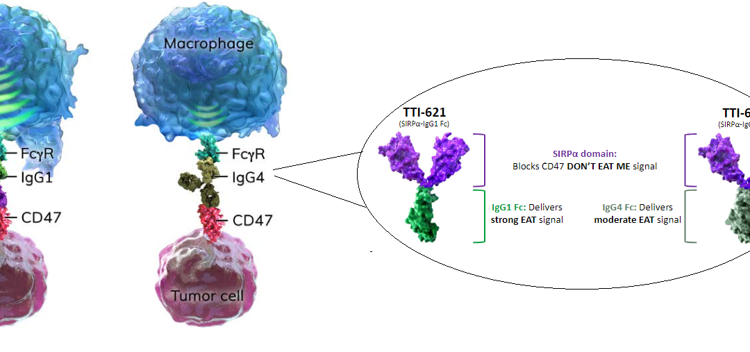
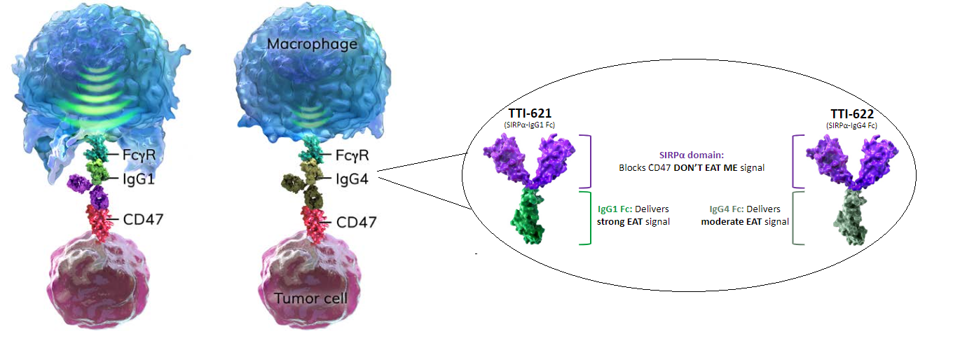
In addition to blocking CD47 through the SIRPα domain, companies are also adding “eat me” signals to enhance the anti-tumor activity. Companies are using different types of antibody isotypes, mainly IgG1 and IgG4, to trigger anti-tumor activity.

Below is a visual representation of a macrophage, representing the immune system defense mechanism, and the CD47 blocking agents that activate the immune system to detect tumor cell.

  • FCγR: FC receptor found on surface of innate cells
  • IgG1 and IgG4: antibody isotypes that deliver the “eat me” signals
  • CD47: “do not eat me” signal found on surface of tumor cell

Gilead’s $4.9 Billion Acquisition of FourtySeven Validated CD47 Space

In early 2020, Gilead struck a deal to buy development stage company FortySeven for $4.9 billion. The takeover gave Gilead control of Phase 1 anti-CD47 antibody magrolimab. FourtySeven had reported 15/39 (38%) overall response rate (ORR) in Diffuse large B-cell lymphoma (DLBCL) patients that had received >3 lines of treatment. Importantly, the response rate was for a combination of FourtySeven’s magrolimab + rituximab. This acquisition kickstarted the interest in CD47 space.

Trillium’s Data Has Best in Class Potential

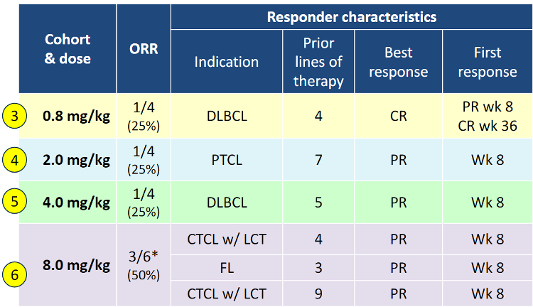
The data that piqued big pharma and healthcare fund interest came from Trillium’s latest cohort reported last week. Cohort 6 (8mg/kg) in relapsed/refractory lymphoma showed 3/6 patients (50%) experienced a partial response (PR). This data was superior to lower dosed Cohorts 3-5 (see below) which had response rates of 25%.

This is an important event for Trillium because these responses are in heavily pre-treated patients (with at least 3 prior lines of therapy) and demonstrate monotherapy activity across a broad range of lymphoma indications. In all Cohorts tested to date, Trillium has shown that 6/18 patients (33%) saw a response. Importantly there were no serious adverse events. The company is now escalating to 12 mg/kg dose level.

It is difficult to make a head-to-head comparison between Trillium and FourtySeven because (1) Trillium’s data is monotherapy, whereas FourtySeven was in combination with rituximab and (2) the indications are different (TRIL in broad lymphomas, FourtySeven in DLBCL).

The main point of interest in Trillium comes from potential response rates when Trillium’s TTI-622 is combined with existing therapies such as PD-1. Theoretically, if monotherapy TTI-622 shows 25-50% response rates in heavily treated lymphoma patients, combination should be higher. This is, most probably, what drew attention of Pfizer and Wall Street.

Not All CD47 Agents Are Equal

From an efficacy point of view, Trillium’s response rate is promising because it is from a single agent, without added benefits of combination approach (discussed above). Importantly, Trillium has reported a relatively clean safety profile.

Although it is overexpressed on tumor cells, CD47 is also expressed in many normal cells, including red blood cells and platelets.  This exposes CD47 antibodies to induce side effects like red blood cell clumping (“hemagglutination”) and anemia, both seen with FourtySeven’s Phase 1 study. Trillium’s candidates, on the other hand, have shown not to bind to red blood cells, which reduces risk of anemia and other blood related adverse events.

The next steps for Trillium are to complete their Phase 1 dose-escalation studies and move on to Phase 2 studies that target specific hematology indications. The good news is that Trillium will have nearly $300 million to advance their pipeline forward.